Energy Resolved Neutron Imaging (ERNI) at FP5


About Flight Path 5 (ERNI)
Flight Path 5 (ERNI) utilizes thermal neutrons from the 1L target, providing neutrons from 1 meV to 1 keV for nuclear physics measurements and ~10 meV to 100 eV for energy-resolved neutron imaging. Flight paths from 6 meters to 60 meters are available and allow a variety of experiments, e.g. imaging with beam spot sizes from 1mm to ~1m in diameter. The flight path is primarily used for neutron imaging/tomography, especially in energy-resolved mode, but can also be used for detector development. Many samples characterized by energy-resolved neutron tomography on flight path 5 are also characterized with neutron diffraction on SMARTS or HIPPO. Together with cold neutron imaging at Asterix and high energy neutron imaging at Target 4’s 60R beamline, Flight Path 5 complements the proton radiography at LANSCE and high energy x-ray radiography offered by AET-6, providing LANL with a complete and unique set of CT and radiography capabilities.
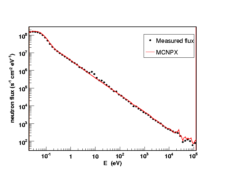
The measured and calculated neutron flux for 100 uA running at Flight Path 5, with a flight path length of 9.73 meters (nominal placement of the fission chamber).
Instrument specifications
Flight Path 5 (ERNI) has two experimental locations: A cave in ER1 of the Lujan Center, covering source-detector distances from ~6.5 m to 11m, and a station accessible by a silo for source-detector distances of 58-62m. The two stations are connected by an evacuated 45 m long pipe through the vulcanic rock. Variable collimation allows beam spots from mm to several cm in the cave to almos 1 m in the silo. A 3,000 channel time-of-flight imaging detector with 512x512 pixels over 28x28 mm2 is the workhorse for energy-resolved neutron imaging. The ability to record ~3,000 neutron radiographs, each at a different neutron energy, allows imaging contrast across neutron energies from cold to epi-thermal. For isotopes with neutron absorption resonances between 1 and 100 eV, 3D isotope distribution or isotope density can be measured non-destructively.
All of this can be done with materials in containers, i.e. nuclear fuels or weapons materials. Using radiographs recorded at neutron energies where materials with high thermal neutron absorption are transparent, energy-resolved neutron imaging also allows to characterize materials opaque to thermal neutrons such as plutonium specimen. The imaging capability is also applied to study fossils for which X-ray CT methods can not be applied, e.g. larger specimen or fossils with iron rich minerals. Studies of water-uptake in plants utilize the large contrast that hydrogen atoms in water provide for neutron radiography.
Examples
Characterization of nuclear fuels and actinide materials
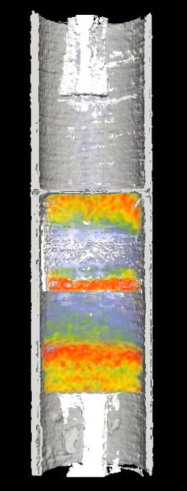
Five urania fuel pellets in a steel rodlet. Two of the pellets were sintered in an RF furnace while sitting on a tungsten disk resulting in some tungsten diffusion into the pellets. While high thermal neutron attenuation shows steel and hydrogen containing glue, weak thermal neutron attenuation shows voids. The tungsten density, measured by neutron absorption resonances, is visualized in color and ranges from 0 to 1 atom % relative to the uranium (collaboration with the LANL Accident Tolerant Fuel program, Kenneth J. McClellan and Andy Nelson).
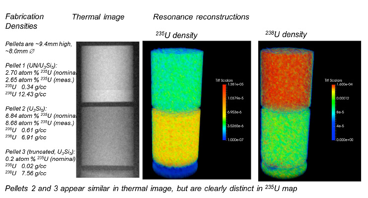
Measurement of enrichment levels in nuclear fuels and 3D measurement of isotope densities (collaboration with the LANL Accident Tolerant Fuel program, Kenneth J. McClellan and Andy Nelson).
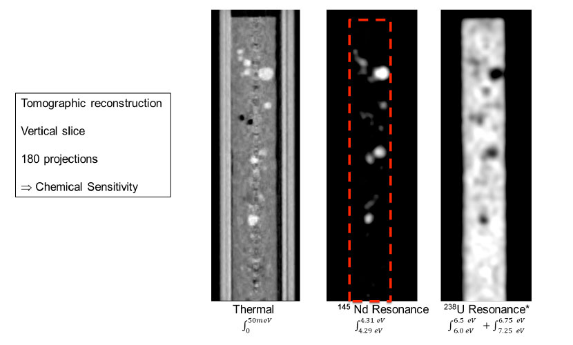
Visualization of rare-earth inclusions, including Nd, in a surrogate transmutation nuclear fuel (collaboration with nuclear fuel programs at LANL and Idaho National Laboratory).
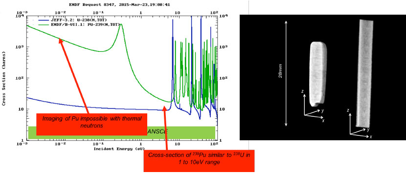
Tomographic reconstruction of Pu-Ga alloy sample. The samples are opaque to thermal neutrons, but can be studied with the ability to select neutron energies greater than thermal energies, where these materials are transparent (with Alice Smith, MST-16).
Tomographic reconstruction of an additively manufactured tungsten test object
Close inspection of such CT scans allows to characterize manufacturing flaws non-destructively, complementing characterization with LANSCE diffraction instruments such as HIPPO and SMARTS (with Michelle Espy & James Hunter, AET-6).
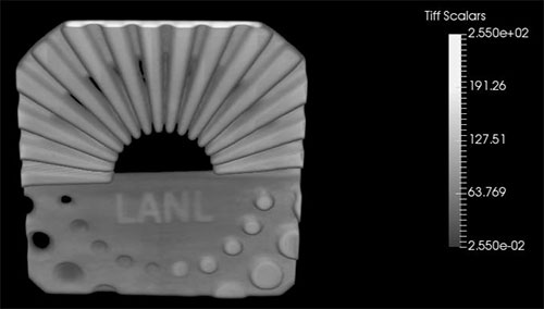
Tomographic reconstruction of an additively manufactured tungsten test object.
Remote sensor-less partial pressure measurements in gases
Using neutron absorption resonance analysis, the pressure in gases can be determined. For a Xe gas contained in a steel container with a pressure gauge reading of 758 kPa, the pressure determined from neutron resonances was 739 kPa. Since this method is isotope specific, the partial pressure of gases in gas mixtures can be determined, e.g. to characterize fission gases in a spent or irradiated fuel (collaboration with A. Tremsin, UC Berkeley).
Element specific imaging
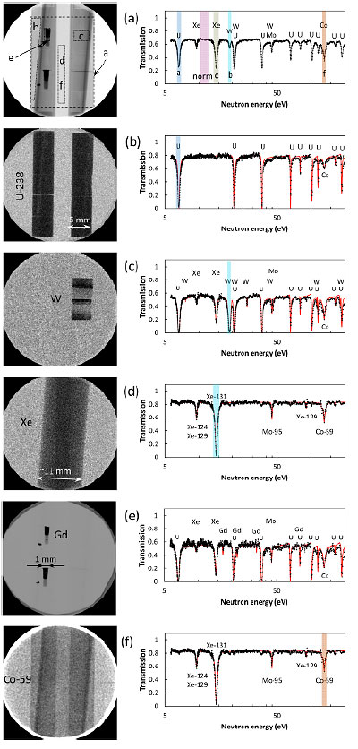
Element-specific images. (a) Full spectrum (white beam) radiograph and a spectrum obtained in the area indicated by a wide dashed rectangle. The letters in the radiograph indicate the areas for which spectra are extracted in figures (b)-(f). The energy ranges shown by the shaded areas in the spectrum of figure (a) indicate the energy range for the subset of images which were used to reconstruct the element-specific maps (b)-(f), except for the map of Gd, where thermal neutrons were used due to high absorption cross section of Gd. 238U, W, Xe and Gd are imaged separately from other elements despite the fact they were overlapped with each other as well as with steel pipes. Steel pipes are visualized by the resonance of 59Co in image (f), which is normalized by the full spectrum open beam image. Iron itself has non-negligible attenuation in the entire range of measured energies, but does not have resonances. (with A. Tremsin, UC Berkeley)
Water transport in plants
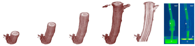
The high contrast provided by neutrons for water allows to study water uptake in living trees. The light areas in the tomography indicate water content. The last two radiographs on the right show a tree standing in a cup of D2O with a NMR coil around the stem. The 2nd radiograph is a difference picture where green areas show the highest change in D2O content, indicating that the center of the stem does transport water (LANL LDRD, S. Sevanto of EES-14).
Optimization of crystal growth parameters in scintillators
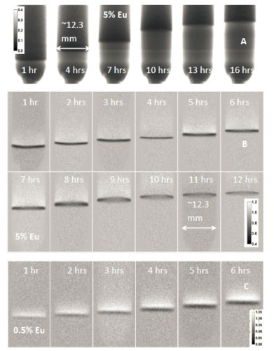
Upscaling of promising scintillator systems to commercial production levels is hampered by identifying the ideal growth conditions in a Bridgman furnace for a given system. Parameters like temperature profile and growth speed affect the yield (e.g. due to cracking of materials with anisotropic distribution of the dopants) and performance (e.g. due to dopants concentrated in small volumes). Energy-resolved neutron imaging is a unique in situ tool, which can characterize dopant distributions and solid/liquid interfaces in situ (collaboration with DOE/NA-22 funded venture lead by E. Bourret-Courchesne/LBNL)
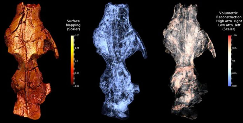
Characterization of fossils
The availability of X-ray CT scanners has revolutionized the field of paleontology. However, in some cases the layers of rocks are too thick for conventional x-rays or certain minerals, e.g. iron rich minerals, prevent the use of conventional X-ray and neutrons can make a contribution. In a collaboration with T. Williamson’s group at the University of New Mexico in Albuquerque and the New Mexico Museum of Natural History, fossils of mammals and dinosaurs were studied at LANSCE. The example below shows the CT reconstruction of surface as well as weak and strong neutron attenuation of part of a skull of Tetraclaenodon Puercensis, a herbivorous mammal that lived in New Mexico 60-63 million years ago. The weak neutron attenuation shows channels, such as the root of the teeth.